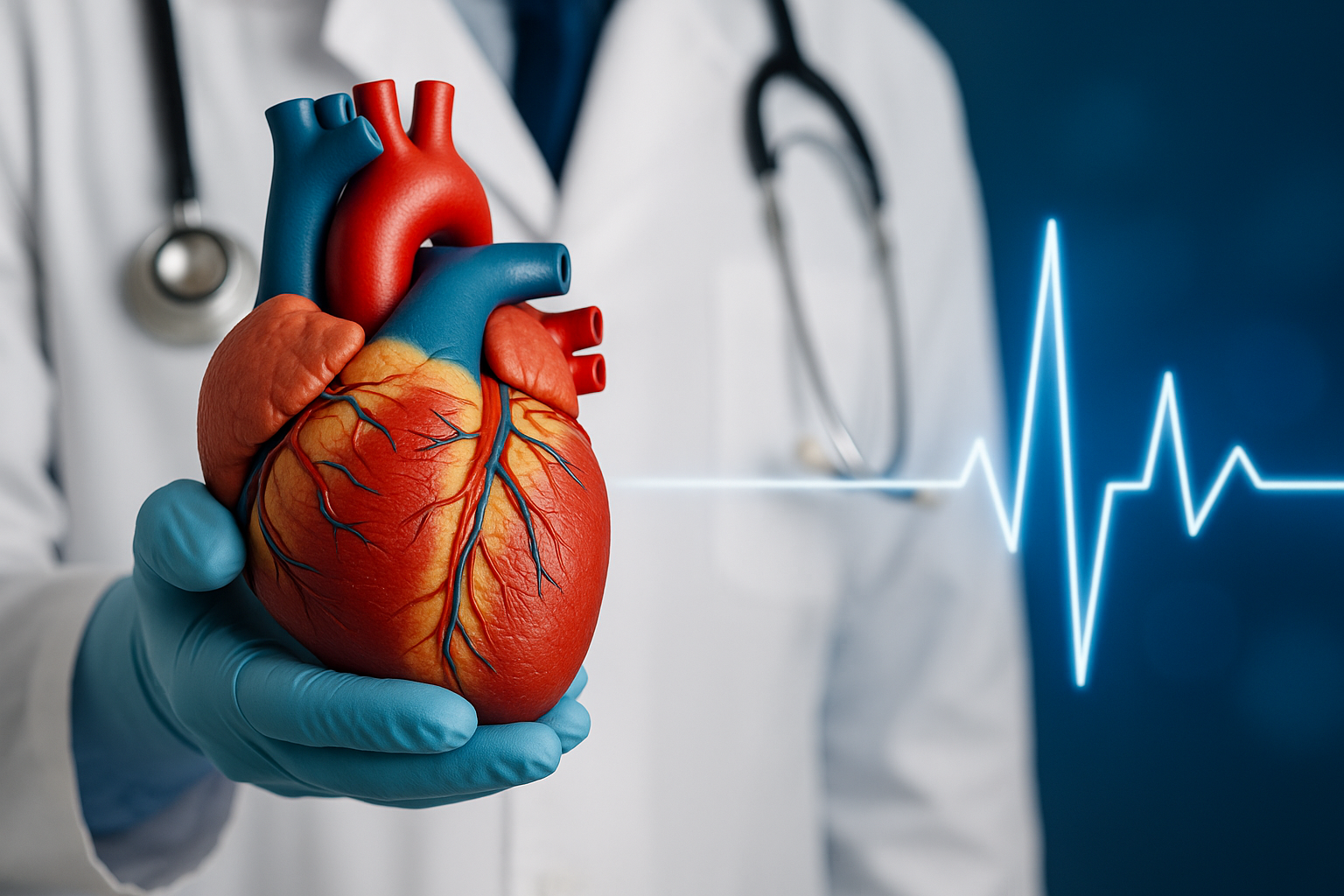
Cardiovascular Surgery
- WHAT IS CARDIOVASCULAR SURGERY AND WHICH CONDITIONS DOES IT TREAT?
- HOW ARE CORONARY ARTERY DISEASE AND BLOCKED VESSELS TREATED SURGICALLY?
- HOW ARE HEART VALVE DISEASES DIAGNOSED AND TREATED?
- WHAT IS CAROTID ARTERY SURGERY AND WHEN IS IT NECESSARY?
- WHAT IS AORTIC ANEURYSM SURGERY AND HOW IS IT PERFORMED?
- HOW ARE HEART RHYTHM DISORDERS TREATED SURGICALLY?
- WHAT ARE THE RISKS, RECOVERY PROCESS, AND FOLLOW-UP AFTER CARDIOVASCULAR SURGERY?
Cardiovascular surgery is a medical specialty focused on diagnosing, treating, and surgically correcting diseases that affect the heart and blood vessels. These conditions often develop silently over many years and may eventually lead to life-threatening complications if left untreated. Cardiovascular surgeons treat structural problems of the heart, narrowing or blockage of blood vessels, rhythm disorders, and serious conditions such as aortic aneurysms.
This field covers a wide range of diseases, including coronary artery disease, heart valve disorders, carotid artery stenosis, congenital structural abnormalities, and advanced circulatory problems. Depending on the patient’s condition, treatment may involve minimally invasive procedures, open-heart surgery, the use of grafts or artificial valves, or endovascular techniques.
Early diagnosis, appropriate imaging, and timely surgical intervention significantly improve survival and long-term quality of life. At OFM Hospital, our cardiovascular surgery team evaluates each patient with a multidisciplinary approach, combining advanced technology, modern surgical methods, and personalized treatment plans.
The overall goal of cardiovascular surgery is to restore healthy circulation, improve heart function, prevent future cardiovascular events, and enhance the patient’s overall well-being.
Coronary artery disease occurs when the heart’s blood vessels become narrowed or completely blocked, preventing sufficient oxygen-rich blood from reaching the heart muscle. If these blockages progress, they can lead to chest pain, heart failure, rhythm disturbances, or even a heart attack. Surgical treatment becomes necessary when medications or less invasive interventions such as angioplasty and stent placement cannot adequately restore blood flow.
The most established surgical method for treating advanced coronary artery disease is Coronary Artery Bypass Grafting (CABG). During this procedure, the surgeon uses a healthy blood vessel—often taken from the leg, chest, or arm—to create a new pathway that bypasses the blocked artery. This allows oxygenated blood to reach the heart muscle without obstruction. CABG is especially effective for patients with multiple blocked vessels, diabetes-related vascular disease, or severe left main artery stenosis.
Modern surgical techniques allow coronary bypass operations to be performed with or without the heart-lung machine, depending on the patient's condition. Off-pump (beating-heart) surgery can minimize complications in suitable patients, while on-pump surgery may be preferred for more complex cases requiring precise reconstruction.
Proper preoperative evaluation—including coronary angiography, echocardiography, blood tests, and anesthetic assessment—is critical for determining the safest and most effective surgical approach. After surgery, patients typically recover in the intensive care unit for close monitoring before transitioning to the ward for gradual mobilization and breathing exercises.
The goal of surgical treatment is to restore adequate blood flow, relieve symptoms such as chest pain, reduce the risk of heart attack, and improve long-term survival. When performed at a modern medical center like OFM Hospital, coronary bypass surgery has high success rates and significantly enhances the patient’s quality of life.
Heart valve diseases occur when one or more of the heart’s valves fail to open or close properly, disrupting normal blood flow. These conditions may develop due to aging, congenital abnormalities, infections such as rheumatic fever, or degenerative changes in the valve structure. Over time, valve dysfunction can lead to shortness of breath, fatigue, swelling in the legs, fainting, irregular heartbeats, or heart failure if not treated appropriately.
Diagnosis begins with a detailed clinical evaluation, including physical examination and cardiac imaging. Echocardiography (ECHO) is the primary tool used to assess valve function, measure blood flow across the valve, and determine whether the problem is stenosis (narrowing) or regurgitation (leakage). Additional tests such as ECG, chest X-ray, CT angiography, or cardiac MRI may be used to evaluate the heart’s structure and plan the most suitable treatment.
Treatment options depend on the severity of the valve disorder and the patient’s overall health. Mild cases may be managed with medication and regular monitoring, but moderate to severe valve disease often requires surgical intervention. The two main surgical techniques are valve repair and valve replacement. Valve repair aims to preserve the patient’s own valve by reshaping, tightening, or reinforcing the valve structure, and is generally preferred when anatomically feasible. Valve replacement, on the other hand, involves implanting a mechanical or biological prosthetic valve when repair is not possible.
Modern valve surgery can be performed using minimally invasive methods through smaller incisions, reducing recovery time and postoperative discomfort. In eligible patients, newer techniques such as transcatheter valve replacement (TAVR/TAVI) offer alternatives without the need for open-heart surgery.
After the procedure, close follow-up is essential to monitor heart function, check prosthetic valve performance, adjust medications, and prevent complications such as valve thrombosis or infection. With appropriate treatment and regular postoperative care, most patients experience significant improvement in symptoms and long-term quality of life.
The carotid arteries are the major blood vessels located on each side of the neck that supply oxygen-rich blood to the brain. Over time, these arteries may become narrowed due to the buildup of plaque, a condition known as carotid artery stenosis. This narrowing significantly increases the risk of stroke, especially when plaque becomes unstable or when blood flow is critically reduced.
Most patients with carotid artery disease do not notice symptoms until the condition becomes advanced. Some individuals may experience warning signs known as transient ischemic attacks (TIAs), which include temporary weakness, numbness, difficulty speaking, or vision loss lasting only a few minutes. These episodes signal that the brain is not receiving enough blood and that immediate medical evaluation is necessary.
Diagnosis typically involves Doppler ultrasound, which measures blood flow in the carotid arteries. In more advanced cases, CT angiography or MR angiography may be required to precisely evaluate the location and severity of the blockage. Treatment decisions depend on the degree of narrowing and the patient’s overall stroke risk.
When the narrowing becomes severe—generally above 70%—or when symptoms such as TIA occur, carotid artery surgery (carotid endarterectomy) is often recommended. During this procedure, the surgeon removes the plaque buildup from the inside of the artery, restoring normal blood flow to the brain and significantly reducing the risk of future stroke. In certain cases, carotid artery stenting may be used as a minimally invasive alternative for patients with anatomical or medical conditions that make surgery high-risk.
Carotid surgery is a highly effective preventive procedure when performed at experienced centers. After the operation, patients are closely monitored for blood pressure control, neurological status, and wound healing. Long-term follow-up includes lifestyle changes, medical therapy, and periodic imaging to ensure the artery remains open.
By intervening early and managing risk factors such as smoking, diabetes, high blood pressure, and high cholesterol, the likelihood of severe stroke can be dramatically reduced. Carotid artery surgery plays a crucial role in safeguarding brain health and protecting patients from life-threatening complications.
An aortic aneurysm is a dangerous enlargement or weakening of the aorta—the body’s largest artery responsible for carrying oxygen-rich blood from the heart to the rest of the body. When a portion of the aorta dilates beyond its normal diameter, the vessel wall becomes fragile and prone to tearing or rupturing. A ruptured aortic aneurysm is a life-threatening emergency, making early diagnosis and timely surgical treatment essential.
Aortic aneurysms are typically categorized based on their location: thoracic aortic aneurysms, which occur in the chest region, and abdominal aortic aneurysms, which occur below the diaphragm. Most patients do not experience symptoms until the aneurysm becomes large. In some cases, individuals may report chest pain, abdominal discomfort, back pain, or a pulsating sensation in the abdomen. Routine imaging—such as abdominal ultrasound, CT scan, or MRI—is crucial for early detection, especially in patients with risk factors like smoking, high blood pressure, advanced age, or a family history of aneurysm.
The decision to pursue surgical treatment depends on the aneurysm’s size, growth rate, and the patient’s overall condition. Generally, aneurysms larger than 5–5.5 cm, or those that grow rapidly, require intervention to prevent rupture. There are two primary surgical approaches: open aortic surgery and endovascular aneurysm repair (EVAR/TEVAR).
In open surgery, the surgeon replaces the weakened aortic section with a durable synthetic graft, ensuring long-term stability and normal blood flow. Although more invasive, open surgery remains the gold standard for certain complex aneurysms. Endovascular repair, on the other hand, is a minimally invasive procedure in which a stent-graft is inserted through a small incision in the groin and positioned inside the aorta to reinforce the vessel wall. This method allows for faster recovery, shorter hospital stay, and reduced postoperative discomfort, making it particularly suitable for high-risk patients.
After surgery, patients require careful monitoring in the intensive care unit, followed by gradual mobilization and long-term follow-up imaging to ensure the graft remains stable and properly functioning. Lifestyle modifications such as smoking cessation, blood pressure control, and regular cardiovascular check-ups are critical to preventing complications and protecting overall vascular health.
Aortic aneurysm surgery plays a vital role in preventing life-threatening rupture and significantly improves both survival rates and long-term quality of life when performed at an experienced cardiovascular center like OFM Hospital.
Heart rhythm disorders, also known as cardiac arrhythmias, occur when the electrical signals that coordinate the heartbeat function abnormally. These irregularities may cause the heart to beat too fast, too slowly, or inconsistently, leading to symptoms such as palpitations, dizziness, fatigue, chest discomfort, and shortness of breath. While many arrhythmias can be managed through medication or catheter-based procedures, certain complex or persistent conditions require surgical intervention.
Surgical treatment is most commonly recommended for patients with atrial fibrillation (AFib), particularly when the arrhythmia significantly affects quality of life or fails to respond to medical and minimally invasive therapies. One of the most effective surgical techniques is the Maze procedure, during which precise incisions or controlled energy applications are made in the heart’s atria to create new electrical pathways. These pathways guide electrical impulses correctly, restoring a stable heart rhythm. The Maze procedure can be performed as open-heart surgery or as a minimally invasive operation through smaller incisions.
In some cases, arrhythmias develop due to structural abnormalities or scarring within the heart tissue. Surgical removal of the problematic tissue or reconstruction of the heart structure may be necessary to prevent recurrent rhythm disturbances. Additionally, certain rhythm disorders may require implantation of pacemakers or implantable cardioverter defibrillators (ICDs) when the heart’s natural electrical system fails to maintain a safe rhythm.
Preoperative evaluation is crucial to determine the type of arrhythmia and identify its underlying cause. Diagnostic tests often include ECG, Holter monitoring, electrophysiological studies, cardiac imaging, and blood tests. These assessments help the surgical team decide whether the patient will benefit more from surgery, catheter ablation, or hybrid procedures combining both methods.
Following surgery, patients require close monitoring to assess rhythm stability, adjust medications, and ensure effective healing. Most individuals experience a significant improvement in symptoms and a reduced risk of stroke, heart failure, and long-term complications associated with uncontrolled arrhythmias. With modern techniques and experienced surgical teams, rhythm surgery is a safe and successful option for restoring normal heart function.
Every cardiovascular surgery carries specific risks, but with modern technology, experienced surgical teams, and advanced perioperative care, most patients undergo these procedures safely and with excellent long-term outcomes. Understanding the potential risks and the expected recovery process helps patients prepare mentally and physically for the journey ahead.
Common risks associated with cardiovascular surgery include bleeding, infection, heart rhythm disturbances, temporary kidney dysfunction, and reactions related to anesthesia. Although these complications are uncommon, the surgical team takes extensive precautions to minimize them. High-risk patients—such as those with diabetes, advanced age, severe lung disease, or previous heart surgery—receive personalized preoperative assessments to determine the safest approach.
The recovery period typically begins with close monitoring in the intensive care unit (ICU), where vital signs, heart rhythm, and oxygen levels are carefully observed. As soon as the patient stabilizes, they move to the ward for continued recovery. During this phase, breathing exercises, early mobilization, and proper pain management are essential to prevent complications such as pneumonia or blood clots. Nurses and physiotherapists guide patients through progressive activity plans to support healthy healing.
Once discharged, patients are advised to follow a structured home-care routine, including medication management, wound care, walking programs, and dietary adjustments. Follow-up appointments at OFM Hospital often include physical examinations, ECG, echocardiography, blood tests, and additional imaging when necessary. These check-ups allow the medical team to monitor heart function, evaluate graft or valve performance, and ensure the patient is recovering as expected.
Lifestyle modifications—such as quitting smoking, maintaining a healthy weight, engaging in regular physical activity, and controlling blood pressure, diabetes, and cholesterol—are critical for long-term success. Many patients benefit from cardiac rehabilitation programs that provide supervised exercise and education to support recovery and reduce the risk of future heart complications.
With proper postoperative care and long-term follow-up, most individuals experience significant improvements in symptoms, physical capacity, and overall quality of life. The goal of cardiovascular surgery is not only to treat existing disease but also to help patients return to safe, active, and fulfilling lives.
Specialist Dr. Mustafa Kar
Specialist Dr. Mustafa Kar
Erciyes University Faculty of Medicine
Medical Education
Ankara Dışkapı Training and Research Hospital
Residency in Cardiovascular Surgery
Konya Training and Research Hospital
Cardiovascular Surgery Specialist
Ankara Maltepe Yaşam Hospital
Cardiovascular Surgeon
Alanya Can Hospital
Cardiovascular Surgeon
Hayat Heart Hospital
Cardiovascular Surgeon
Memorial Antalya Hospital
Senior Cardiovascular Surgeon
Private OFM Hospital, Antalya
Cardiovascular Surgery Department
Department: Cardiovascular Surgery
Education: Erciyes University Faculty of Medicine
E-mail: mustafa.kar@ofmantalya.com
Languages: English
-
Coronary Artery Bypass Surgery
-
Heart Valve Replacement
-
Carotid Artery Surgery
-
Varicose Vein Treatment
